Procedures
Hip Arthroscopic Surgery

Experiencing pain in the hip
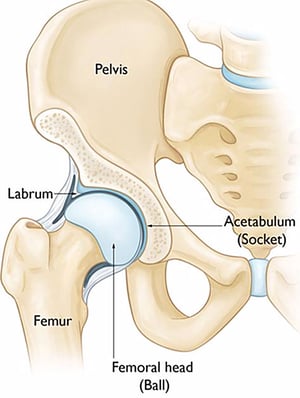
Please seek medical help if you have any of these symptoms. Your orthopaedic surgeon will take a detailed medical history from you, perform a thorough medical examination and likely organise an MRI scan of your hip to confirm the labral tear.
FAI femoral acetabular impingement
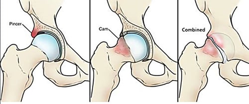
The CAM type of FAI occurs due to an extra bump of bone at the junction of the femoral head and neck. This extra bone collides or impinges on the labrum when the hip is moved around. Imagine having a small stone in your shoe! The soft labrum is therefore squeezed between the bony acetabulum and bony bump and ends up tearing. If the bony bump is not removed and the labrum is not repaired, the hip will become arthritic sooner than later.
The pincer type of FAI occurs when there is overhanging or extra bone around the acetabulum. As the hip is moved, the pincer crushes the soft labrum against the hard femoral neck, resulting in a badly damaged labrum and painful hip. Some patients have a combined CAM and pincer which makes their symptoms worse.

Arthroscopic labral repair
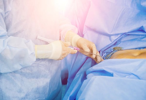
Many years ago, repair of the labrum involved open surgery where a large skin incision was made. These days arthroscopic or keyhole surgery is performed for the same purpose. Small incisions allow wounds to heal faster, lower the risk of infection and reduces the collateral damage to other structures in the hip. Several incisions about 1 or 2cm long are made around your painful hip. The instruments used for this procedure are long and thin like chopsticks. It allows accurate localisation of the torn labrum and precise repair of the tear. Labral tears should also not be repaired in isolation. The offending CAM bony bump or overhanging pincer must be addressed to prevent repeat tears in future.
Rehabilitation after surgery may take 3 to 6 months. In the early days after surgery, medication is given to manage general pain around the hip, ice is applied to reduce swelling and patients are not allowed to bend their hips beyond 90 degrees to protect the repair site. While the arthroscopic scars are small and heal in 2 weeks, patients need to go for frequent physiotherapy after surgery to prevent scarring in their hips, maintain muscle strength in the thighs and promote smooth a walking pattern. Cycling on a stationary bicycle is important to get your hips going to prevent stiffness. Most patients return to brisk walking at about 3 months and sports at 6 months.
The intention of hip preservation surgery is to repair and patch up the hip injuries and delay the need for a formal hip replacement. There is a chance that damage in the hip is too advanced and patients continue to have pain despite hip preservation surgery. In these cases, we would counsel patients for a total hip replacement so that they may return to their active lifestyles again. It is not true that only elderly patients receive a total hip replacement and it is not true that you cannot be active after hip replacement surgery. Do speak to us if you have been bothered by your hip pain. We are equipped with a spectrum of non-surgical and surgical techniques to help you with your symptoms.
What is hip arthroscopy, and what conditions does it treat?
Hip arthroscopy is a minimally invasive, keyhole procedure in which a surgeon inserts a small camera and fine instruments through tiny 1 to 2cm incisions to diagnose and treat problems inside the hip joint, without a large open incision.
It is commonly used to treat:
- Labral tears, where the cartilage rim of the socket is repaired and stitched back
- Femoroacetabular impingement (FAI), by removing abnormal fragments of bone
- Loose fragments of bone or cartilage within the joint
- Certain cartilage injuries and inflammation of the joint lining
Keyhole techniques help reduce the risk of infection and collateral damage to other structures in the hip, and are associated with minimal blood loss. They are most suitable for younger, active patients with non-arthritic hip conditions.
What are the most common causes of hip pain?
Hip pain can arise from the joint itself or from the surrounding structures. Common causes include:
- Labral tears: damage to the cartilage rim of the socket, often causing groin pain with clicking or clunking sensations
- Femoroacetabular impingement (FAI): excess bone along the edges of the socket or femoral neck that causes the labrum to tear and cartilage to wear
- Avascular necrosis (AVN): disruption of the blood supply to the femoral head, which can lead to collapse and arthritis
- Hip arthritis: degenerative wear and thinning of the cartilage, causing pain, stiffness, and a limp
Because hip and groin pain can also be referred from the lower back, an accurate diagnosis by a hip specialist is essential before starting treatment.
What is hip preservation surgery, and how does it help avoid a replacement?
Hip preservation surgery uses arthroscopic (keyhole) techniques to rectify and treat injuries around the hip and prevent or delay the need for a formal hip replacement. If simple injuries of the hip are left untreated, they may escalate into full arthritis over time.
Preservation techniques include:
- Arthroscopic labral repair: torn labrum is stitched back to the rim of the socket through small incisions
- Arthroscopic pincer and CAM resection: abnormal fragments of bone from FAI are removed using burr and shaver instruments
- Core decompression and bone graft: for early-stage AVN, to relieve pressure in the femoral head and encourage healing
- Intra-articular injections: hyaluronic acid injections to improve pain and function in early hip arthritis
These techniques aim to treat the underlying problem early and keep your natural hip joint for as long as possible.
How is hip arthroscopy different from hip replacement?
The two procedures address different problems and patient groups:
- Hip arthroscopy: a keyhole procedure that preserves and repairs the natural hip joint, best for younger patients with labral tears, impingement, or loose bodies. It is not used for advanced arthritis.
- Hip replacement: replaces the worn joint with an implant, best for older patients or those with advanced arthritis and significant cartilage loss
In short, arthroscopy repairs the existing joint, while replacement substitutes one which is beyond repair. Your surgeon will recommend the right option based on your age, activity level, diagnosis, and the degree of joint damage seen on imaging.
What is the recovery time after hip arthroscopy?
Recovery from hip arthroscopy is generally quicker than from open hip surgery, but it still requires a structured rehabilitation programme:
- First 1 to 2 weeks: walking with crutches to protect the joint while pain and swelling settle
- Weeks 2 to 6: progressive physiotherapy to restore movement and strength
- 6 to 12 weeks: return to most daily activities and light exercise
- 3 to 6 months: return to sport and higher-impact activity, depending on the procedure performed
A formal rehabilitation program that patients embark on after surgery is key to optimising recovery. Your surgeon will tailor the timeline to the specific repair carried out.
Can hip pain be treated without surgery?
Yes. Many causes of hip pain respond well to non-surgical treatment, which is almost always the first line of management:
- Physiotherapy: targeted exercises to strengthen the hip and improve mobility and flexibility
- Activity modification: adjusting movements and reducing high-impact loading
- Weight management: reducing load on the hip joint
- Medication: anti-inflammatories and analgesics for pain relief
- Intra-articular injections: hyaluronic acid and steroid injections are often given to reduce pain in early arthritis
Surgery is considered only when these measures no longer provide relief and the underlying condition warrants it.
How is the cause of hip pain diagnosed?
An accurate diagnosis is the foundation of effective treatment. A hip specialist will typically assess your hip through:
- Clinical history: understanding your symptoms, activity, and how the pain behaves
- Physical examination: testing hip movement, strength, and specific pain triggers
- X-rays: to assess joint space, arthritis, and bone structure
- MRI scan: to evaluate soft tissues such as the labrum, cartilage, and tendons
Because hip pain can be referred from the spine or other areas, a thorough evaluation ensures the true source is identified and the right treatment is chosen.